Prepare the CTA
Normalize orientation and resolution, localize the heart, then crop a 128³ input with the REPRISE-compatible intensity protocol.
Original image stays localORIGINAL V23 · JOINT TPS 600
A three-dimensional image becomes a labelled tetrahedral mesh. One surface network. One shared deformation. Ready to explore.
01 / THE HEART IN MOTION
A complete 20-phase sequence, reconstructed with the released inference pipeline. Each phase starts with its own CTA image.
We screened three complete sequences and selected one with a clear contraction pattern. The animation shows the 20 exported volumetric meshes in acquisition order.
Independent single-phase V23 inference; no temporal filter or interpolated mesh geometry. Playback speed is illustrative.
02 / IMAGE → SURFACE → VOLUME
Normalize orientation and resolution, localize the heart, then crop a 128³ input with the REPRISE-compatible intensity protocol.
Original image stays localThe original V23 network and frozen weights predict nine cardiac surfaces, including the Aorta and pulmonary artery.
No default retrainingGeometric correspondences drive one joint TPS field across all template parts, with Aorta continuation and a tetrahedron determinant guard.
One shared deformation03 / LOOK BENEATH THE SURFACE
Move through the cutaway to see the original tetrahedral elements inside the heart walls. The camera orbits and moves closer; the mesh itself is unchanged.
MMWHS example. The cut removes complete tetrahedra behind a moving plane; no remeshing or display smoothing.
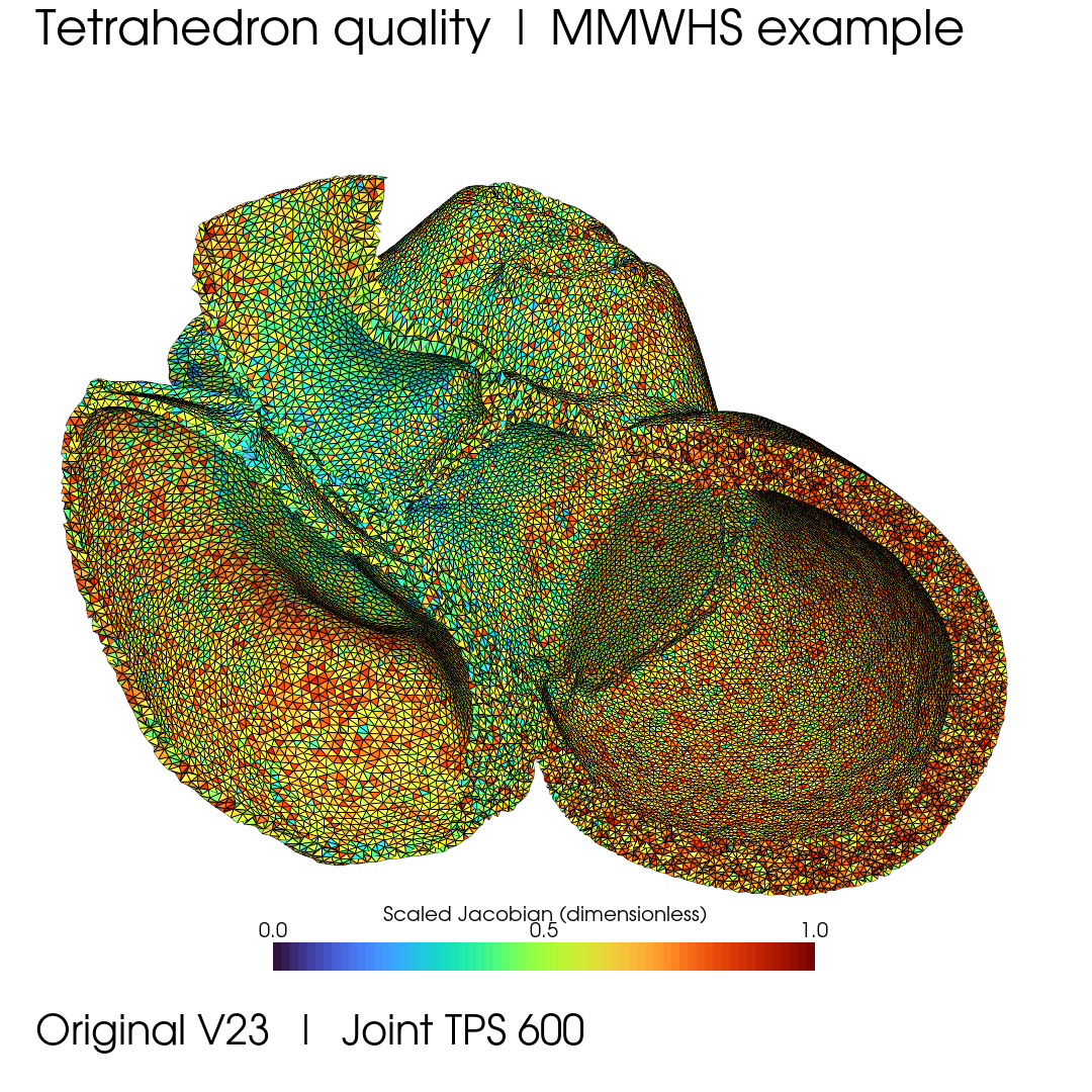
Fengming Turbo-64 · common range [0, 1]. Element quality alone does not establish anatomical or simulation validity.
04 / BEYOND THE DEVELOPMENT DATA
Explore reconstruction examples from MMWHS and ImageCAS with dataset-aware preprocessing and the same released V23 weights.
A labelled whole-heart reconstruction viewed from changing angles. No MMWHS fine-tuning was used for this example.
Native-image preprocessing followed by the original V23 model and shared volumetric deformation. No ImageCAS fine-tuning was used for this example.
These are selected qualitative development examples. The separate 10 MMWHS + 10 ImageCAS surface evaluation is documented in the repository; it is not a 20-case final-volume validation.
05 / MAKE IT YOURS
Run the code directly, or give your coding agent the HeartVolMesh skill. Installation creates an isolated environment and verifies the model and runtime downloads.
Readable source, preprocessing, mesh export, quality reports and visualization scripts.
Explore the GitHub repository ↗A GPU-aware bootstrap and an inference workflow for your own local CTA files.
Install heartvolmesh ↗git clone https://github.com/fmlinks/HeartVolMesh.git
cd HeartVolMesh
python bootstrap.py --workspace ./workspace
python run_inference.py --workspace ./workspace \
--image ./CTA.nii.gz --output ./resultWindows RTX 5090 was tested in a fresh environment. The installer provides a driver-aware RTX 3090 route; that hardware has not been directly tested. Review heart coverage and native CT overlays for each new dataset. This is a research release.